

Large Colon Enterolithiasis Diagnosed with Advanced Imaging and Treated Surgically by Dr. Hugo Almonte
Patient Presentation
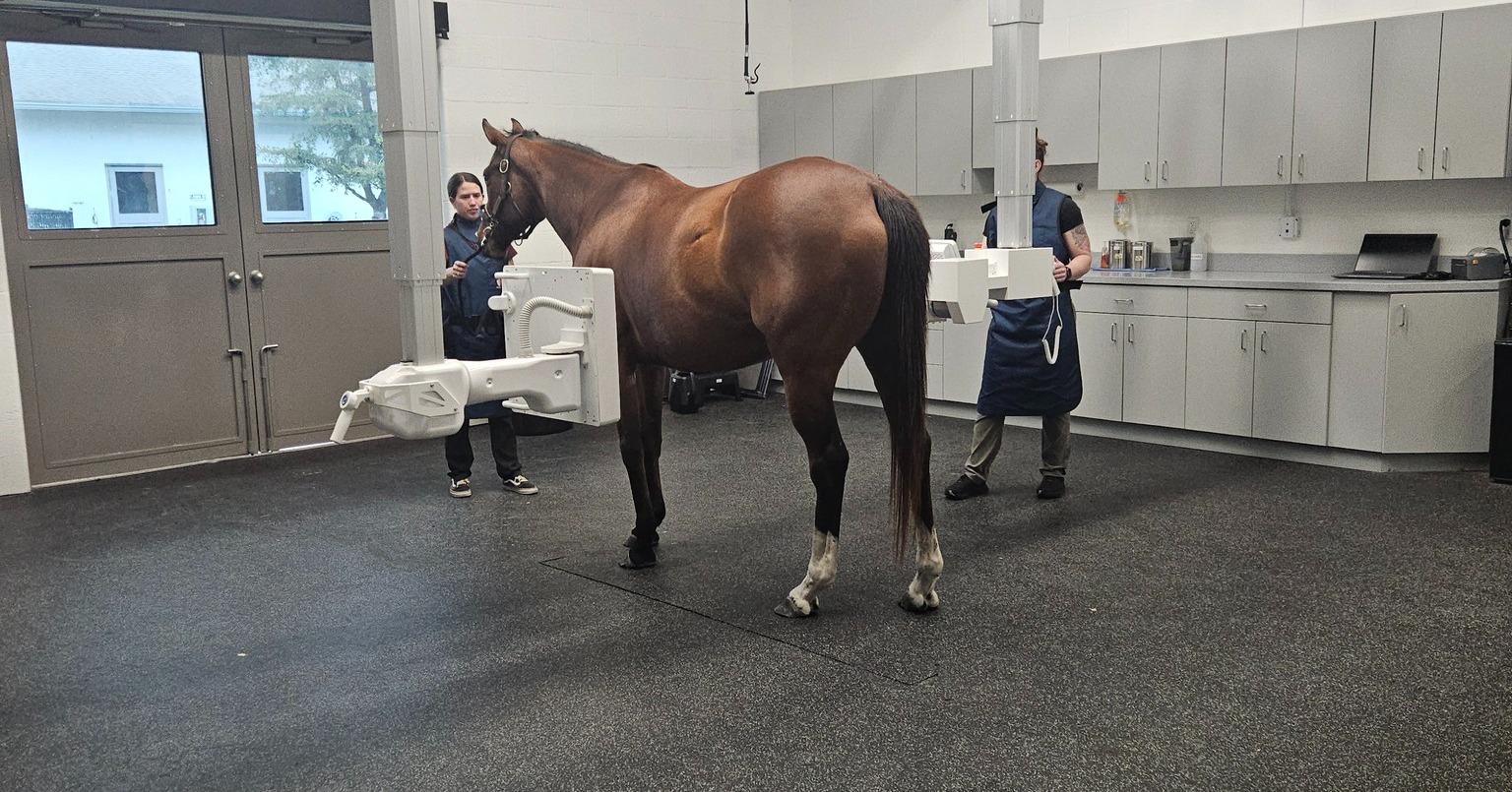
A horse presented as an emergency for clinical signs of colic and was evaluated by the Rood & Riddle Surgery Team.
Initial Evaluation and Findings
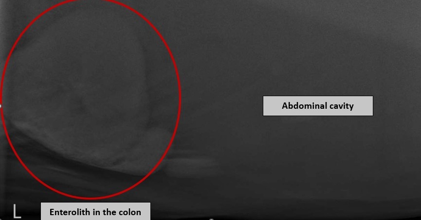
Enteroliths are mineralized concretions that form within the large colon and can obstruct intestinal flow, leading to impaction colic. Their development is multifactorial and may be influenced by diet, breed, geographic region, and soil type, though the exact cause is not fully understood. Diagnostic evaluation included rectal palpation, abdominal ultrasound, and advanced digital radiography. High-powered digital radiographs revealed findings consistent with large colon enteroliths, later confirmed during surgery.
Medical or Surgical Management
Dr. Hugo Almonte performed a routine colic workup and utilized advanced diagnostic imaging to identify the enteroliths. The horse underwent exploratory laparotomy, during which two enteroliths were located and surgically removed from the large colon. Surgical removal is the only definitive treatment for enterolith-associated colic.
Outcome
The enteroliths were successfully removed, resolving the obstruction and addressing the cause of the colic. The horse recovered following surgical intervention with appropriate postoperative care and monitoring.
Educational Takeaway
Enteroliths are an important cause of obstructive colic in horses. Advanced imaging, including high-quality digital radiography, can aid in definitive diagnosis, and timely surgical intervention is essential for a successful outcome in affected horses.
Learn more about Dr. Hugo Almonte: CLICK HERE